
Looking to identify potential skin cancer?
At DermConsult, we recognise just how serious skin cancer can be. As such, we can provide a bespoke consultation for you to identify the different types of skin cancer and provide suitable treatments and procedures.
There are two main types of skin cancer: melanoma and non-melanoma. The main non-melanoma skin cancers are basal cell carcinomas and squamous cell carcinomas.
Basal cell carcinoma (BCC) is the commonest type of skin cancer in the UK. BCCs rarely metastasise. Thus, although it is a skin cancer, it almost never poses a threat to life.
A squamous cell carcinoma (SCC) is a type of non-melanoma skin cancer. It is the second most common type of skin cancer in the UK. SCCs can invade local tissues and may sometimes metastasise. SCCs can be life-threatening.
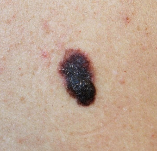
Melanoma is a potentially life-threatening type of skin cancer that arises from the uncontrolled growth of melanocytes (skin pigment cells). It is more likely than other types of skin cancer to metastasise.
Occasionally the diagnosis is clear from the appearance of the lesion. A skin biopsy (sample) under local anaesthetic can confirm a diagnosis.
The treatment of a BCC depends on its type, size and location. Most BCCs are removed surgically. Options include:
Diagnosis of a SCC is usually made by a dermatologist. The appearance and story behind the lesion give clues that point towards a SCC. A biopsy (sample) or excision (complete removal) under local anaesthetic can confirm the diagnosis; a histopathologist will closely examine the tissue under a microscope.
There may also be further investigations to determine whether the SCC has metastasised. These include imaging (such as CT and MRI scans) and lymph node (gland) biopsies.
Treatment differs on a case-by-case basis. If the SCC is just local, surgery is usually the best treatment option. This will involve removing the SCC with some normal skin around it to reduce the chances of any cancer being left behind. Radiotherapy (firing X-rays at the lesion) is an alternative to surgery. If the SCC has spread beyond the initial site, a combination of treatments is often required; surgery, radiotherapy and chemotherapy may all be required to treat the SCC in this scenario.
Melanoma may be suspected because of the features of the lesion such as its appearance or a history of change in an existing mole. A dermatologist uses an instrument named a dermatoscope to look more closely at the suspicious area of the skin.
The best way of diagnosing a melanoma is to completely excise (surgically remove) it, as well as a small surrounding margin of normal skin tissue. A histopathologist will then carefully study the specimen under a microscope and confirm and characterise the melanoma.
If the melanoma is more advanced, imaging (such as CT and MRI scans) and lymph node biopsies (samples) may be needed to investigate whether the cancer has metastasised.
Following the diagnostic excision, a second surgical procedure named a wide local excision is recommended. This involves removing a larger margin of tissue (typically 1cm-2cm extra) from around the former melanoma site. This will reduce the chances of any cancer cells being left behind which could cause recurrence or even spread of the melanoma. Depending on the microscopic thickness of melanoma, a procedure named sentinel lymph node biopsy may be discussed. Opinions are divided about this investigation which is only offered in some centres. It is a tool in determining how a melanoma is likely to behave (risk of recurrence) after it has been treated but does not make any difference in the survival rate.
In more advanced melanoma, radio- and chemotherapy may be amongst the supplementary therapies needed to treat cancer metastases. Targeted genetic therapies and immunotherapy are also powerful tools in combating advanced melanoma. In addition, dermatology centres often run clinical trials which test new types of treatment. Many recent advances in treatment are promising
If you are suffering from skin cancer, simply get in touch and book your consultation with our friendly team. At DermConsult we’re committed to providing the very best quality care and a personalised service for each and every one of our patients. Our experienced consultant dermatologists have a dynamic and forward-thinking approach, applying the best recent evidence when diagnosing and treating their patients.